Stanford's Super-Resolution Microscopy Unveils SARS-CoV-2 Replication in Unprecedented Detail
June 1, 2024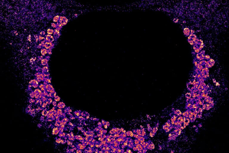
A Stanford University team has developed a new super-resolution microscopy technique to observe the SARS-CoV-2 virus replicating in cells at the nano-scale level.
The technique involves labeling viral RNA with fluorescent molecules and using a blinking method to capture high-resolution images.
These images reveal how the virus forms spherical structures around the cell nucleus with unprecedented detail.
This method enhances drug development precision by allowing nanoscale study of virus replication and cell processes.
The team plans to use the technique to study the effects of antiviral drugs on viral replication and to map all 29 proteins of the SARS-CoV-2 virus.
This breakthrough can significantly advance virology research and improve our understanding of viral infections.
Summary based on 1 source
Get a daily email with more Science stories
Source
Phys.org • May 31, 2024
A new way to see viruses in action: Super-resolution microscopy provides a nano-scale look